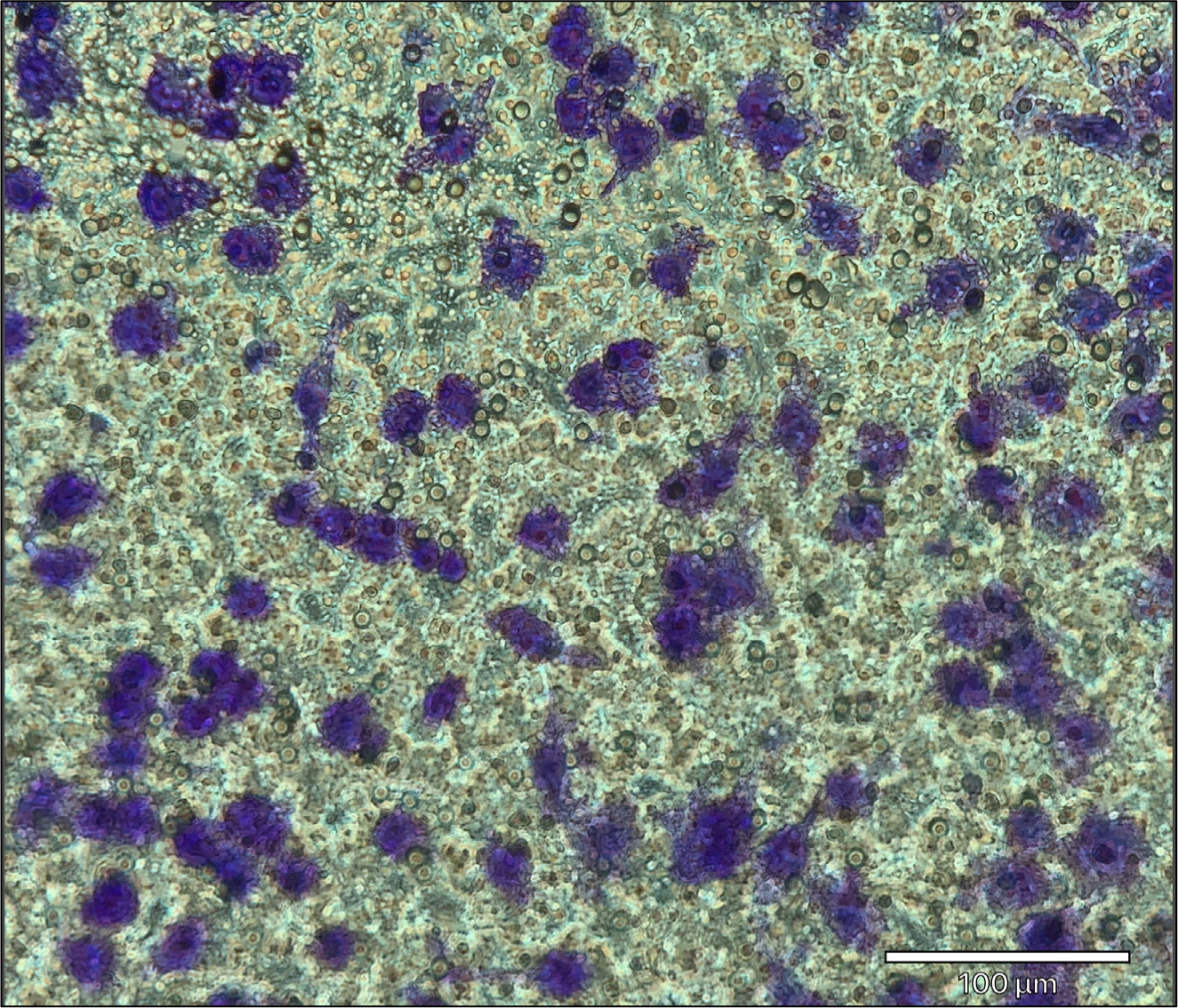
Figure 3.

Representative image of migrated cells stained with crystal violet. J774A.1 macrophage/monocyte migration using 100 ng/mL recombinant mouse C5a. Migrated cells are stained with 0.2% crystal violet solution. Images of the transwell membrane are taken with a 20x objective using the ECHO Revolve 3 microscope. Migrated J774A.1 cells are stained purple. Scale bar = 100 μm.
