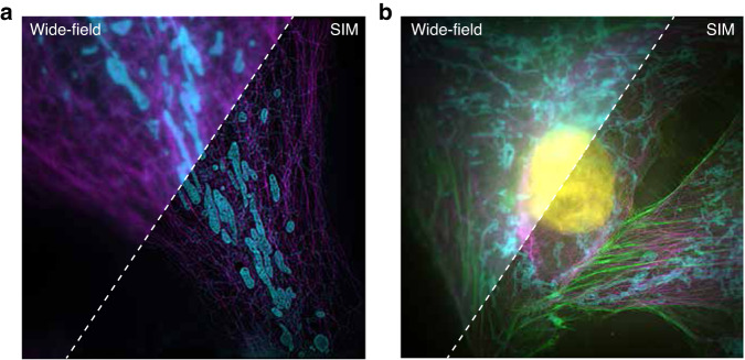
Fig. 1. Comparison of a wide-field image and SIM image obtained using the Airy Polar-SIM system200.
a Two-color (561-PK mito RED labeled mitochondria (cyan) and 640-SiR-tubulin kit—labelled tubulin (magenta)) imaging results of homemade sample COS7 cells. Please refer to Appendix 1 for the specific production process. b Four-color (DAPI-labelled nuclei, yellow; Alexa 647-labelled tubulin, magenta; Alexa 555-labelled actin, green; Alexa 488-labelled mitochondria, blue) imaging results of fixed cells. The sample was purchased from Standard Imaging Co. Ltd

