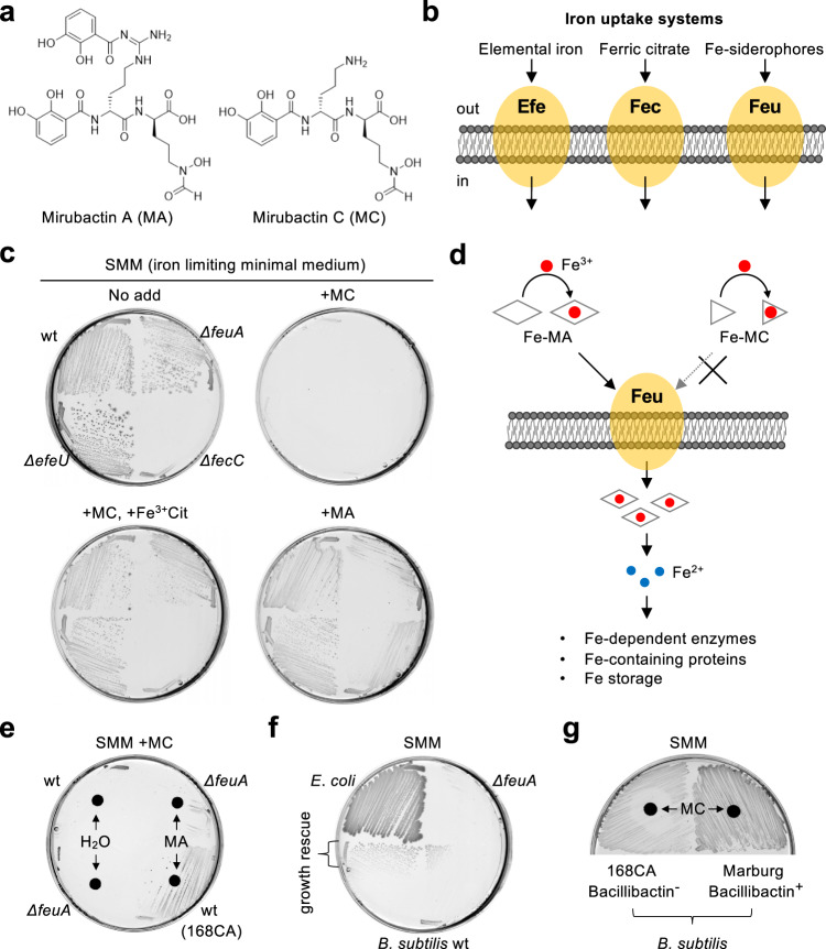
Fig. 1. Mirubactin C inhibits iron uptake and its utilization.
a Schematic representation of structures of Mirubactin A (MA) and C (MC). b Schematic representation of iron uptake systems in B. subtilis. c Growth inhibition by MC in iron-limiting minimal medium (SMM). B. subtilis strains 168CA (wild-type), YK2739 (ΔefeU), YK2740 (ΔfecC) and YK2741 (ΔfeuA) were streaked on SMM plates with or without 20 μg/ml MA, 10 μg/ml MC and/or 100 μM ferric-citrate (Fe3+Cit), and incubated for 30–42 h at 37 °C. d Schematic representation of effects of Mirubactins on iron uptake and utilization. e Uptake and utilization of iron in B. subtilis mediated by MA. 168CA (wild-type) and YK2741 (ΔfeuA) were streaked on a SMM plate in the presence of 10 μg/ml MC, with paper disc containing 6 μl of H2O or 20 mg/ml MA, and incubated for 30–42 h at 37 °C. f Uptake and utilization of iron in B. subtilis mediated by E. coli. E. coli strain BW25113 and B. subtilis strains (wild-type 168CA and ΔfeuA) were streaked on a SMM plate containing 10 μg/ml MC, and incubated for 30–42 h at 37 °C. g Growth of B. subtilis Marburg strain in the presence of MC. 168CA (bacillibactin-) and Marburg (bacillibactin+) strains were streaked on a SMM plate with paper disc containing 6 μl of 5 mg/ml MC, and incubated for 30–42 h at 37 °C. The figures are representative of at least three independent experiments.

