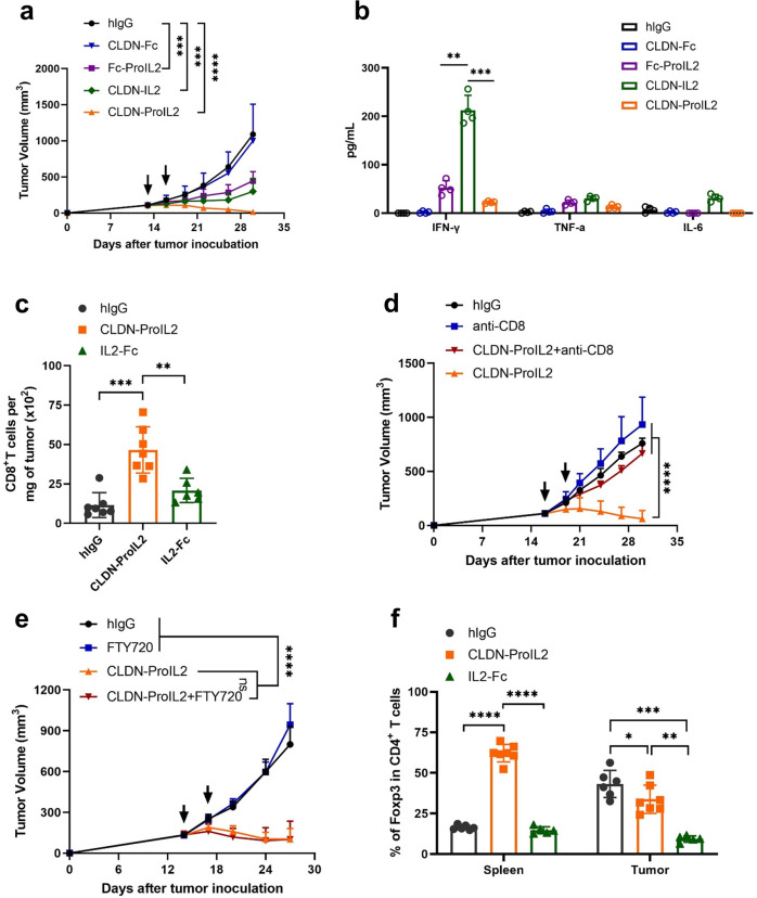
Fig. 1.
CLDN-ProIL2 targets CTLs inside TME while increases Treg cells in the peripheral with reduced toxicity and enhanced antitumor efficacy. a, b C57BL/6 J mice (n = 10/group) were subcutaneously inoculated with 5 × 10⁵ MC38-CLDN and treated with hIgG, CLDN-ProIL2 (60 μg) and equimolar doses of CLDN-Fc, Fc-ProIL2 or CLDN-IL2 by intraperitoneal injection on days 13 and 16 post-tumor inoculation. Tumor volume was measured twice a week (a). Serum was collected and isolated at 24 h post the first dose treatment. Cytometric Bead Array was used to quantify the amount of serum IFN-γ, TNF-α and IL-6 (b). c, f C57BL/6 J mice (n = 10/group) were subcutaneously inoculated with 5 × 10⁵ MC38-CLDN and treated with hIgG, CLDN-ProIL2 (60 μg) and equimolar doses of IL2-Fc by intraperitoneal injection on days 13 and 16 post-tumor inoculation. Four days after the second treatment, splenocytes and TILs were analyzed for the absolute number of CD8+ T cells per milligram of tumor tissues (c) and the frequency of Foxp3+CD4+ T cells (f). d MC38-CLDN tumor-bearing C57BL/6 J mice (n = 10/group) were treated with hIgG or CLDN-ProIL2 (60 μg) on days 15 and 18. Mice were intraperitoneally treated with anti-CD8 (200 μg/mouse) twice a week starting on day 14. Tumor volume was measured twice a week. e MC38-CLDN tumor-bearing C57BL/6 J mice (n = 10/group) were treated with hIgG or CLDN-ProIL2 (60 μg) on days 14 and 17. To block T cells migrating from lymph node into the tumor site, mice were administered with FTY720 every 2 days starting on day 13, and through the end of the experiment. Tumor volume was measured twice a week. Data are shown as means ± SEM. a–f is a pool of two independent experiments. The P value was determined by two-way ANOVA with Geisser-Greenhouse correction (a, d and e), one-way ANOVA with Tukey’s multiple comparisons test (b, c and f). * P < 0.05, ** P < 0.01, *** P < 0.001 and **** P < 0.0001, ns not significant

