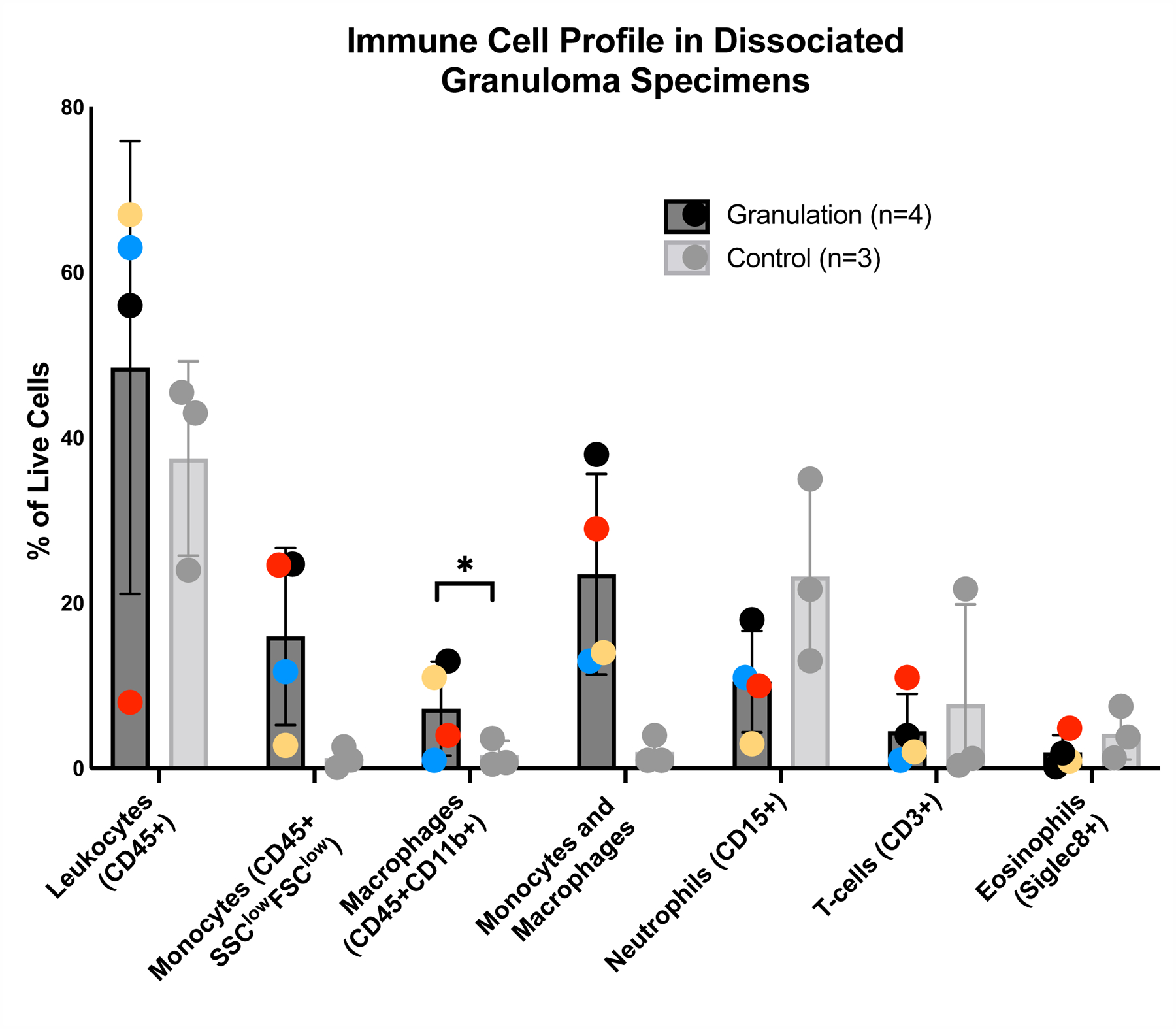
Figure 2: Immune cell profile in dissociated tracheal granulation tissue.

Dissociated granulation tissue and control samples were stained and analyzed using flow cytometry to identify leukocytes (CD45+), macrophages (CD45+CD11b+), monocytes (CD45+FSClowSCClow), including classical CD14highCD16low, intermediate CD14highCD16high, non-classical CD14lowCD16high) distinguished using forward and side scatter properties, T-cells (CD3), neutrophils (CD15), and eosinophils (Siglec 8). Error bars signify SEM.
