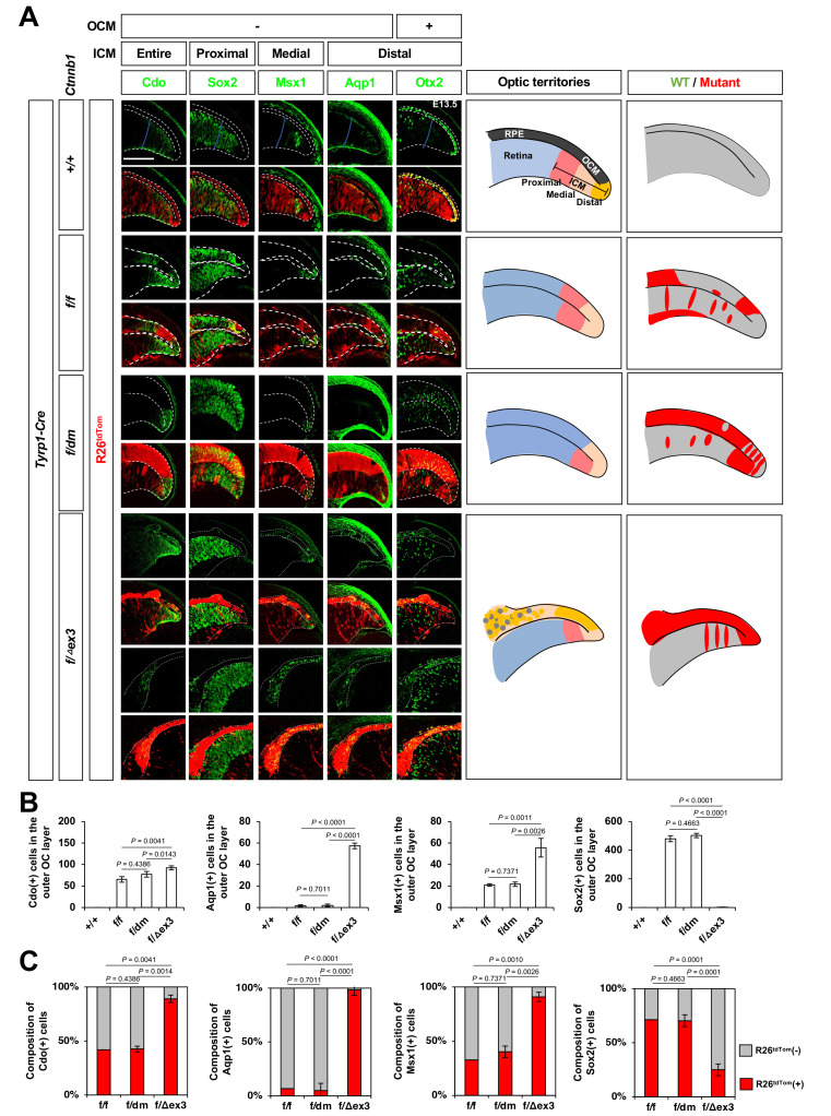
Fig. 2. Ctnnb1 facilitates distal ciliary margin (CM) cell fate in the optic neuroepithelium.
(A) Sections of embryonic day 13.5 (E13.5) Ctnnb1+/+;Tyrp1-Cre, Ctnnb1f/f;Tyrp1-Cre, Ctnnb1f/dm;Tyrp1-Cre, and Ctnnb1f/Δex3;Tyrp1-Cre mouse eyes were stained with the antibodies against Cdo, Sox2, Msx1, Aqp1, and Otx2. Optic neuroepithelial continuum of the retina (inner) and RPE (outer) are marked by the dotted lines. Schematic diagrams depict the peripheral optic cup areas. Scale bar = 50 μm. (B) The cells identified by each cell type-specific marker were counted and the numbers are shown in the graphs. (C) The R26tdTom Cre reporter-expressing cell populations in the marker-positive cells are shown in the graphs. Values in the y-axis are averages and error bars denote the SEM. n = 5, 3 independent litters. OCM, outer ciliary margin; ICM, inner ciliary margin; RPE, retinal pigment epithelium; WT, wild type; OC, optic cup.

