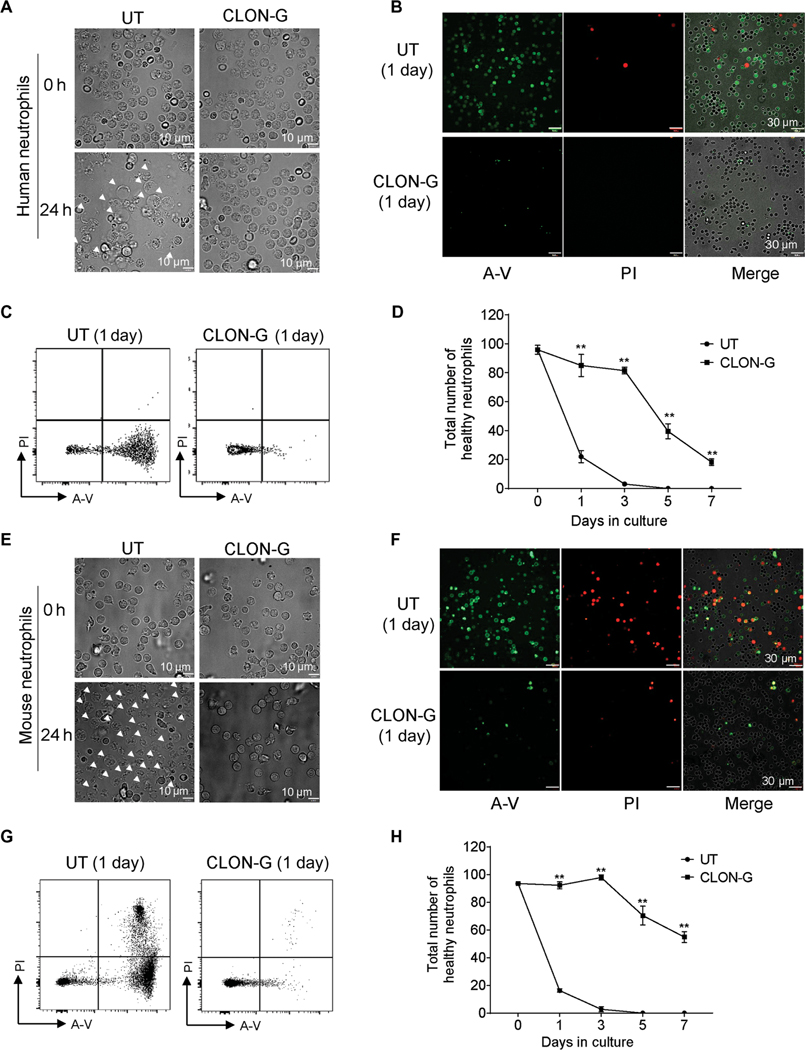
Fig. 1. CLON-G, a treatment that simultaneously targets multiple death mechanisms, delays neutrophil death, increasing neutrophil half-life from less than 1 day to greater than 5 days.
(A) Freshly isolated human peripheral blood neutrophils were cultured in RPMI 1640 with 20% FBS with or without CLON-G. The morphologies of untreated (UT) or CLON-G–treated (CLON-G) cells were observed after 1 day by light microscopy. White arrowheads indicate dying cells. (B and C) Neutrophils were stained with FITC–annexin V (A-V; green) and PI (red) after culturing for 1 day. Cell death was assessed by confocal fluorescence microscopy (B) and flow cytometry (C). (D) The total number of healthy neutrophils was calculated in untreated and CLON-G– treated cell populations at the indicated time points as the number of remaining cells (intact cells in fig. S3A) multiplied by the proportion of double-negative cells by flow cytometry (shown in fig. S3C). (E) The morphologies of untreated or CLON-G–treated murine neutrophils were observed after 1 day by light microscopy. White arrowheads indicate dying cells. (F and G) Murine neutrophils were stained with FITC–A-V and PI after culturing for 1 day. Cell death was assessed by confocal fluorescence microscopy (F) and flow cytometry (G). (H) The total number of healthy neutrophils was calculated in untreated and CLON-G–treated cell populations at the indicated time points. All data are presented as means ± SD of three experiments. **P < 0.001 compared to the corresponding untreated group.

