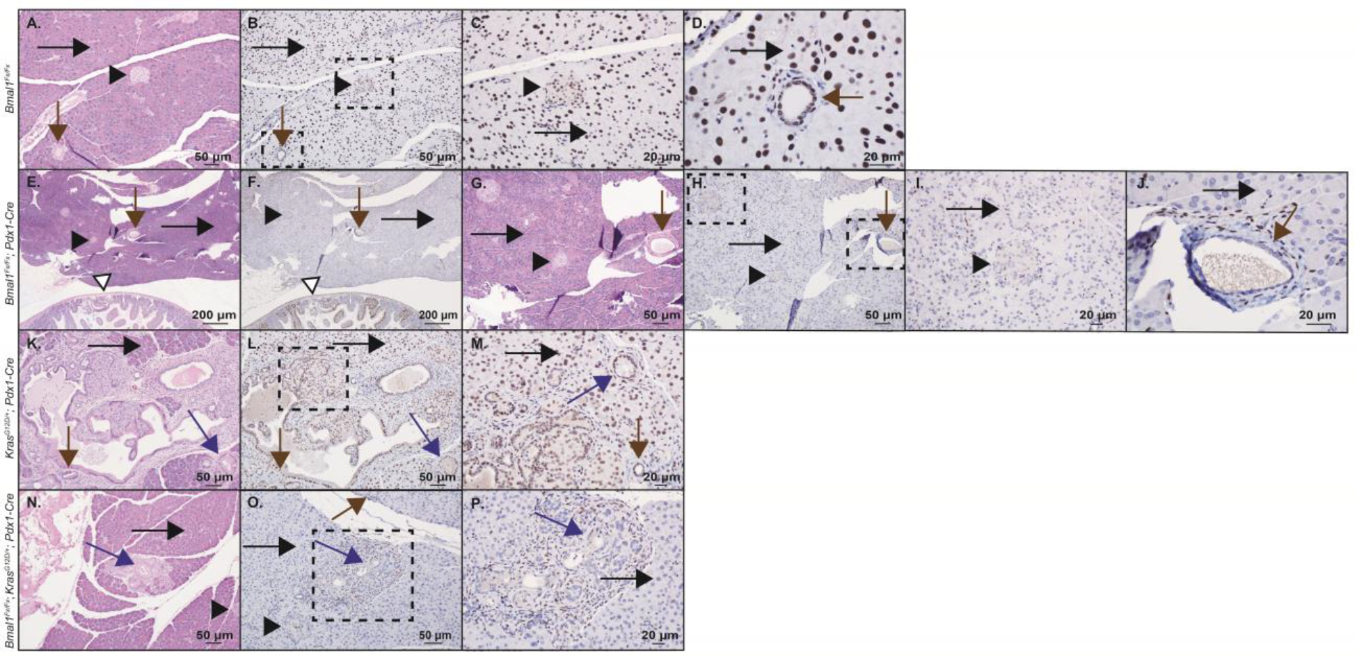
Figure 5: Bmal1 protein expression in pancreas.

Shown are H&E staining and immunohistochemistry (IHC) staining, including high power fields for BMAL1 protein in Bmal1fx/fx mouse pancreas (wild-type control) (A-B, 100x; C, 200x; D, 400x), Bmal1fx/fx; Pdx1-Cre pancreas (E-F, 40x; G-H, 100x; I, 200x; J, 400x), KrasG12D/+; Pdx1-Cre pancreas (K-L, 100x; M, 200x), and Bmal1fx/fx; KrasG12D/+; Pdx1-Cre pancreas (N-O, 100x; P, 200x). In each of the panels, islet cells are marked with a black arrowhead, acinar cells with a long black arrow, ductal cells with a brown arrow, and PanINs with a blue arrow. In panels (E-F), the duodenal mucosa is also shown as an internal control depicting duodenal mucosal cells that retain BMAL1 expression (white arrow). Dotted boxes indicate the area of subsequent magnification. Scale bars are shown for reference.
