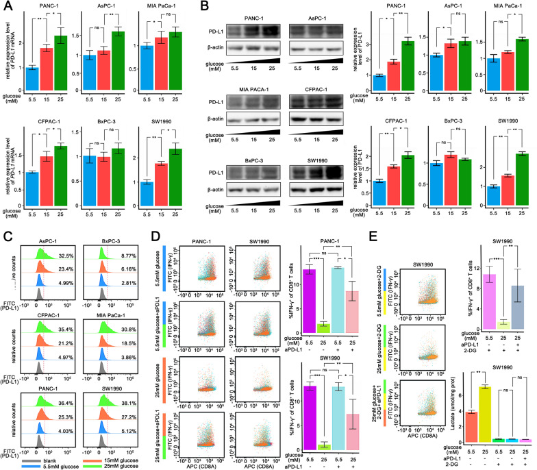
Fig. 2.
Pancreatic cancer cells cultured with high glucose inhibit T cell killing in vitro. A–C qPCR analysis A and Western blotting analysis B and Flow cytometry analysis C of PD-L1 mRNA expression in PANC-1, AsPC-1, MIA PaCa-1, CFPAC-1, BxPC-3 and SW1990 cells 48 h after different sugar concentration (5.5 mM, 15 mM, 25 mM) medium culturing. D Flow cytometry analysis of IFN-γ production of cocultured CD8+ T cells with PANC-1 cells or SW1990 cells (E:T = 1:1) in different treatment. E Flow cytometry analysis of IFN-γ production of cocultured CD8+ T cells with PANC-1 cells or SW1990 cells (E:T = 1:1) cultured in different sugar concentration medium supplemented with 2-DG. PDAC pancreatic ductal adenocarcinoma, TCGA The Cancer Genome Atlas, DC dendric cell, MDSC myeloid-derived suppressor cell, ns non-significant. The graphs show representative results from three independently repeated experiments. *p < 0.05, **p < 0.01, ***p < 0.001

