
Figure 7. Features of inclusions in the rat model confirming α-syn pathology.
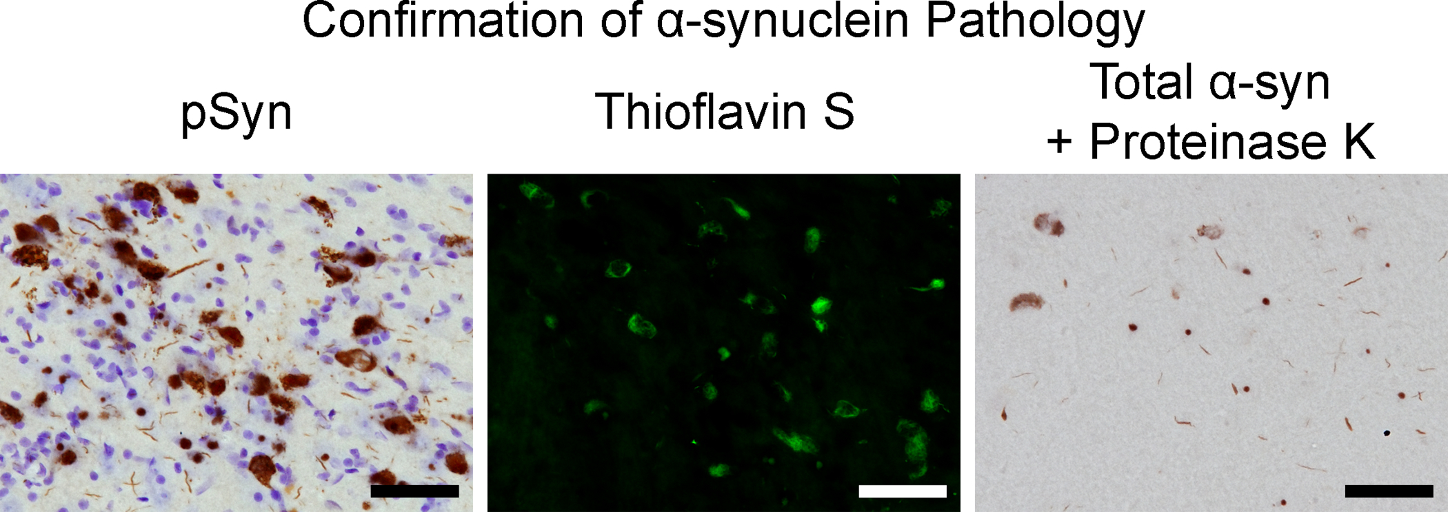
Representative micrographs from the substantia nigra pars compacta at 2 months post-injection. Left: Neurons containing pSyn and counterstained with cresyl violet. Middle: Thioflavin S positive neurons. Right: α-syn-containing inclusions resistant to proteinase K. Scale bar = 50 μm.