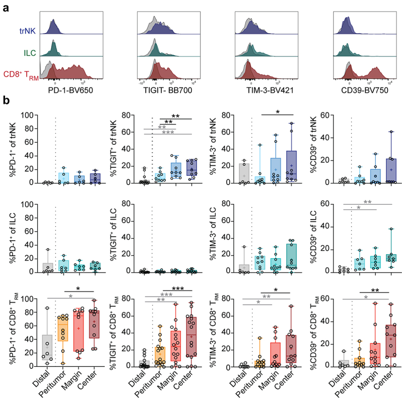
Figure 3.

Immune checkpoint receptor expression on tissue-resident lymphocytes in lung tumors. (a) Representative overlays of expression of PD-1, TIGIT, TIM-3, and CD39 on trNK cells, ILCs, and CD8+ TRM cells in the tumor center. Gray histograms represent FMO controls. (b) Frequencies of trNK cells, ILCs, and CD8+ TRM cells expressing PD-1, TIGIT, TIM-3, or CD39 in different tumor-free tissues or tumor tissues (n = 6–16). Friedman test, Dunn’s multiple comparisons test (patient-matched, black); Kruskal–Wallis test (unmatched, gray). Mean indicated as ‘+’. *p < 0.05, **p < 0.01, ***p < 0.001.
