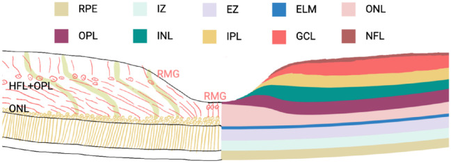
Figure 2.
Two types of Müller cells in the fovea. There are two types of Müller cells in the fovea: specialized Müller cells in the foveola and Müller cells in the foveal walls, which have a characteristic “Z” shape. Müller cells in the foveola are also called Müller cell cones, which run almost straight from the OLM to the ILM. Müller cells in the foveal walls are highly elongated compared to peripheral Müller cells. They run vertically from their foot process in the ILM to the OPL through the Henle fiber layer inward toward the center of the fovea of the retina, where they reach the fovea of the retina almost horizontally and then extend again vertically toward the OLM, forming a characteristic “Z” shape. In this process, obliqued Müller cells and photoreceptor axons constitute the HFL, which is the foveal portion of the OPL. IZ, interdigitation zone; EZ, ellipsoid zone; ELM, external limiting membrane; GCL, ganglion cell layer; NFL, nerve fiber layer.

