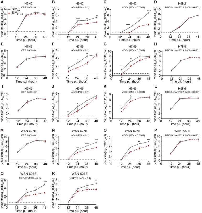
Fig. 8. SIM of NS2 promotes AIV replication in mammalian cells but not in avian cells.
(A to D) Comparison of the replication abilities of H9N2-SIMWT and H9N2-SIME110G virus in indicated cell lines. Primary CEFs (A), A549 cells (B), MDCK cells (C), or MDCK-chANP32A cells (MDCK cells stably expressing chANP32A) (D) were infected with the H9N2-SIMWT or the H9N2-SIME110G virus. Cell supernatants were collected at the described time points post-infection (p.i.), and virus titers were determined using 50% tissue culture infective dose (TCID50) assays. (E to H) Comparison of the replication abilities of H7N9-SIMWT and H7N9-SIME110G virus in indicated cell lines. Experiments were performed as in (A) to (D). (I to L) Comparison of the replication abilities of H5N6-SIMWT and H5N6-SIME110G virus in the indicated cell lines. Experiments were performed as in (A) to (D). (M to R) Comparison of the replication abilities of the avian-signature WSN-PB2-627E virus (WSN-627E, SIMWT) and the avian-signature WSN-PB2-627E virus with NS2 SIM harboring an E110G mutation (WSN-627E, SIME110G) in the indicated cell lines. Experiments were performed as in (A) to (D). In (A) to (R), dotted line denotes the limit of detection based on the dilution factor in our assays. Graphs shown are of one representative (means ± SD, n = 3). Asterisks indicate significant differences between SIMWT and SIME110G, and statistical significance was calculated per time point by unpaired Student’s t test. *P < 0.05, **P < 0.01, ***P < 0.001, and ****P < 0.0001.

