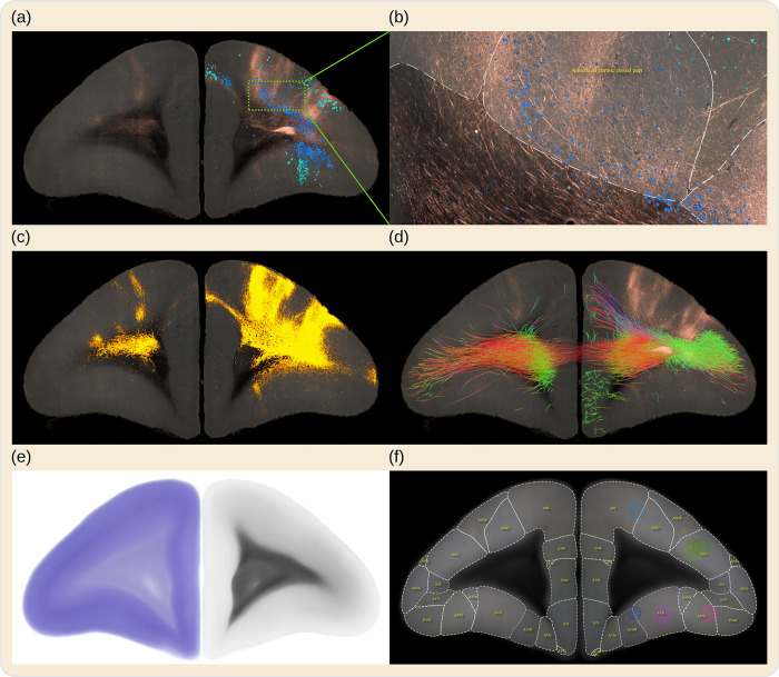
Fig 1. BMCR example images.
The BMCR comprises the image data of 52 anterograde tracer injections and 19 retrograde tracer injections placed into the marmoset PFC, supplemented by retrograde neural tracer data from the Marmoset Brain Connectivity Atlas project (145 injections). This figure shows a series of examples of virtual coronal sections from the BMCR all in the same reference image space. The tracers were injected into Area 8a of the cortex. Panel (a) shows an STPT fluorescent image of the results of an anterograde neural tracer, and as an overlay, neurons labeled by a retrograde tracer dataset from the Marmoset Brain Connectivity Atlas; this dataset originated from a similarly located injection site, mapped to the BMCR image space (the different blue tones, blue and turquoise) indicate whether cells are beneath or above layer IV, information provided by the BMCR. Panel (b) shows a detailed close-up of a portion of the same image. Panel (c) shows the segmented tracer from (a) over the auto-fluorescent background. The overlay in panel (d) shows tractography results of streamlines originating from the same site as the tracer injection, based on averaged dMRI data. The colors reflect streamline directions. The BMCR also includes individual and population average backlit and Nissl images (see (e) for examples of the population average images) and incorporates brain region annotations from major brain atlases for marmosets (see (f) for an example of the Brain/MINDS Atlas). BMCR, Brain/MINDS Marmoset Connectivity Resource; dMRI, diffusion-weighted MRI; PFC, prefrontal cortex; STPT, serial two-photon tomography.

