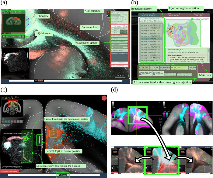
Fig 4. Interaction between BMCR-Explorer and Nora-StackApp.
Interface of the BMCR-Explorer and the Nora-StackApp. Each example shows 2 anterograde tracers in the BMCR reference space. (a) The BMCR-Explorer shows high-resolution microscopy images of neural tracers from different individuals in a common image space. Panel (b) shows the interface for data selection. (c) The cursor position is shown simultaneously in a cortical flatmap and the current coronal section. (d) The Nora-StackApp viewer can show a number of tracer images simultaneously in 3D that facilitates comparative studies. The viewer supports arbitrary virtual sectioning including sagittal, coronal, or transversal sections and can interact with the BMCR-Explorer. The same location can be opened in high resolution in the BMCR-Explorer. Data availability: Panel (a) shows data from 2 marmoset brains with the IDs R04_0079 and R01_0098, panel (c) shows data from R01_0046 and R01_0098, and panel (d) shows data from R04_0080 and R04_0095. The data is publicly accessible from the BMCR-Explorer (http://bmca.riken.jp/), and Nifti stacks can be downloaded from our repository (https://doi.org/10.60178/cbs.20230630-001). BMCR, Brain/MINDS Marmoset Connectivity Resource.

