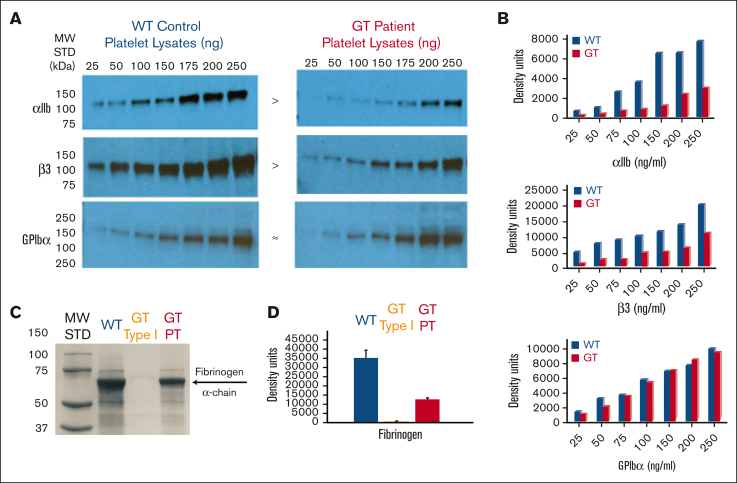
Figure 2.
Quantitative analysis of the patient’s platelet proteins lysed in SDS show reduced integrin αIIbβ3 and fibrinogen levels consistent with variant form of GT. (A) Immunoblot analysis using Abs specific for detection of αIIb, β3, or GPIbα within platelet protein lysates. Increasing quantities of SDS–platelet protein lysates ranging from 25 to 250 ng were loaded into separate lanes of 10% polyacrylamide gels (PAGEs) and electrophoresed under nonreducing conditions. Following transfer to nitrocellulose, the blots were incubated with goat anti-human polyclonal antibodies to αIIb (136 kDa), β3 (125 kDa), or a mAb to human GPIbα (165 kDa). Immunoreactive bands were detected with an horseradish peroxidase–conjugated 2oAb followed by chemiluminescence exposure on radiograph film or a digital imager. The result shown is a representative blot (n ≥ 3 at 7 different lysate concentrations) demonstrating αIIb and β3 appear present at reduced density levels in the GT lysates compared with the WT control, whereas GPIbα appears at similar levels in GT vs WT. (B) Quantitative band density measurements performed with Image J software using photographic film of the immunoblots in panel A exposed to chemiluminescence demonstrate that patient with GT αIIb (30% ± 8%) and β3 (39% ± 11%) is present at reduced levels when comparing the band density with the WT control. As anticipated, the platelet GPIbα levels of patient with GT (94% ± 11%) are nearly identical to a WT control arbitrarily set at 100% in each image. The graphs show MFI in density units for measurements at different concentrations in panel A and converted to percentage ± SEM of WT arbitrarily set at 100% for each representative blot (n = 4). (C) Western immunoblot analysis of platelet protein lysates to detect fibrinogen. Protein (10 μg) was loaded onto a 4% to 20% gradient PAGE and SDS-PAGE performed under reducing conditions. Following transfer to nitrocellulose, the blot was incubated with mouse mAb (5C5) directed against the fibrinogen α-chain (60 kDa). Immunoreactive bands were detected by horseradish peroxidase–labeled m-IgGĸ binding protein, followed by chemiluminescence and detection on photographic film or digital analyzer. As expected, fibrinogen detected in WT platelets was arbitrarily set as the normal level in the (+) control (blue). The apparent absence of fibrinogen in patients with GT type I (αIIbβ3-deficient) platelets served as a (−) control (yellow). Although in stark contrast, appreciable levels were observed in our sample from patient with GT (red). Shown is results of 1 representative blot (n = 4) measured at 3 different exposure times. (D) Fibrinogen band density measurements performed on immunoblot in panel C with Image J software on a scan of the photographic film or digital image used to quantitate the protein level in platelet lysates showed that fibrinogen of patients with GT is present at 34% ± 3% of WT levels of fibrinogen consistent with the levels of αIIb and β3 detected in panel C, while type I GT (−) control had 1.2% ± 0.7% of normal levels. Shown is 1 representative analysis (n = 4 + SEM) measure for at least 3 different exposure times. MW, molecular weight; SDS, sodium dodecyl sulfate; STD, standard.

