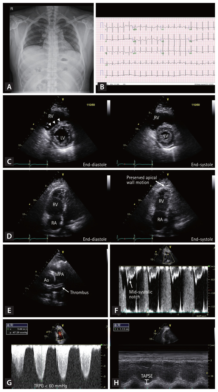
Figure 1.
Typical case of an acute pulmonary embolism (PE) in a 33-year-old man. The initial chest radiography is normal (A). The electrocardiogram demonstrates sinus tachycardia, S wave in lead I (S1), Q wave in the lead III (Q3), T wave inversion in lead III (T3), and precordial leads V1–3 (B). Echocardiography shows D-shaped left ventricle (C, arrowheads), McConnell’s sign (D, arrow), thrombus in the main pulmonary artery (E), mid-systolic notching in the pulsed-wave Doppler tracing of right ventricular outflow tract (F), increased maximal velocity of tricuspid regurgitation (G), and decreased right ventricular contractility assessed by tricuspid annular plane systolic excursion (H). RV, right ventricle; LV, left ventricle; RA, right atrium; Ao, aorta; MPA, main pulmonary artery; TRPG, tricuspid valve peak systolic gradient; TAPSE, tricuspid annular plane systolic excursion.

