
Fig. 1. N-terminal fragments of GSDMB splicing isoforms exhibit different pore-forming activity.
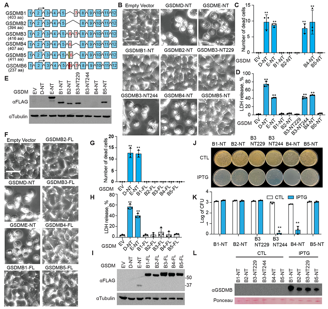
A, Schematic of six alternative splicing variants of GSDMB. Numbered blue boxes represent exons. An alternative splicing acceptor results in an insertion (yellow box) in exon 6 of GSDMB5. The red box in GSDMB6 indicates a 13-nucleotide deletion in exon 6. B-E, The effect of overexpressing GSDMB-NTs on HEK293T cell death, assessed by morphology using microscopy (B and C) and by LDH release (D). Dead cells were counted and quantified using three images (C). Expression of indicated FLAG-tagged GSDMB-NTs was determined by anti-FLAG immunoblot (E). F-I, The effects of overexpressing full-length GSDMBs on HEK293T cell death, assessed by morphology using microscopy (F and G) and by LDH release (H). Dead cells were counted and quantified using three images (C). Expression of indicated FLAG-tagged full-length GSDMBs was assessed by anti-FLAG immunoblots (I). J-L, The effect of overexpressing N-terminal GSDMBs on E. coli cell death, accessed by colony formation on LB plates without (CTL, non-treatment) or with IPTG (J and K). CFU, colony-forming units. Expression of indicated N-terminal GSDMBs was assessed by anti-GSDMB immunoblots (L). Ponceau S stained bands were used as loading controls. Data are mean ± s.d. of biological triplicates and are representative of three independent experiments. Statistical analysis was performed using the two-tailed Student’s t-test. **P < 0.01. Scale bar, 20 μm