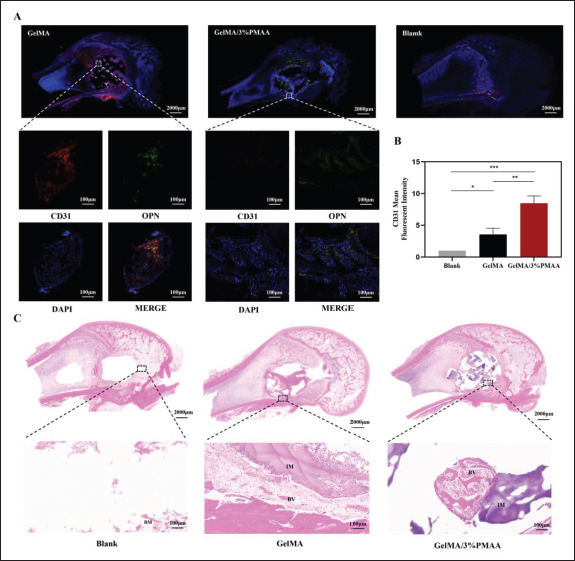
Figure 8.

Histological and immunohistochemistry evaluation of the new blood vessels in the defect site at week 4. (A) Fluorescent staining (CD31 and OPN) of newly formed bone at week 4. (B) The mean fluorescent intensity of CD31. (C) Hematoxylin–eosin staining of newly formed bone at week 4. Data were analyzed using one-way ANOVA and are shown as mean ± standard deviation (*p < 0.05, **p < 0.01, ***p < 0.001, n = 3). (NB, new bone; IM, implanted materials; BM, bone marrow; BV, blood vessel.)
