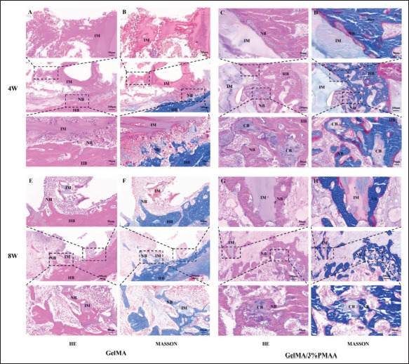
Figure 9.

Histological evaluation of newly formed bone at week 4 and week 8. (A, C) Hematoxylin–eosin (HE) and (B, D) Masson’s trichrome (MASSON) staining in the defect site at week 4. (E, G) HE and (F, H) MASSON staining in the defect site at week 8. (NB, new bone; HB, host bone; IM, implanted materials; CB, chondrogenic bone.)
