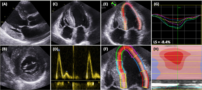
Figure 1: Characteristic echocardiographic features of transthyretin amyloidosis cardiomyopathy.

A: Parasternal long-axis view demonstrating increased interventricular and inferolateral wall thickness; B: Parasternal short-axis view demonstrating increased wall thickness; C: Four-chamber view demonstrating biventricular increased wall thickness; D: Pulsed wave Doppler of the mitral valve demonstrating an E/A ratio >2 and grade 3 diastolic dysfunction; E: Longitudinal strain (LS) measured in the four-chamber view; F: Increased septal apical-to-basal strain ratio; G, H: Longitudinal strain map demonstrating relative apical sparing and a ‘bull’s eye’ strain pattern.
