Fig. 7.
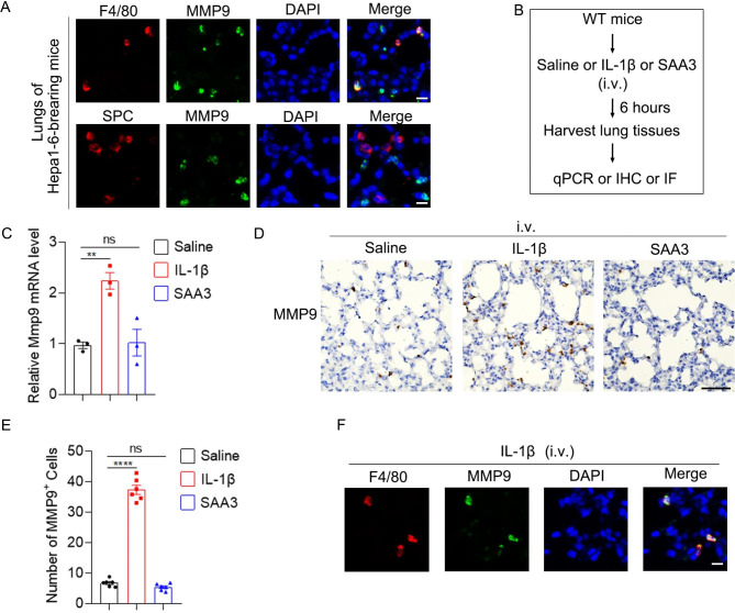
IL-1β increases MMP9 expression in alveolar macrophagesin vivo. (A) MMP9 was expressed by alveolar macrophages but not AT2 cells in the pre-metastatic lung. Double IF staining of F4/80 (red) or SPC (red) and MMP9 (green) in the pre-metastatic lung of Hepa1-6-bearing mice. Scale bar, 10 μm. (B) Schematic for experimental design in C-F. IL-1β (20 ng in 100 ul saline) or SAA3 (1.2 µg in 100 ul saline) were injected intravenously. Six hours later, the lungs were harvested. (C) Intravenous administration of IL-1β but not SAA3 increased MMP9 mRNA level in the lung. (D-E) Intravenous administration of IL-1β but not SAA3 increased MMP9+ cells in the lung. MMP9+ cells were detected by IHC (D). The number of MMP9+ cells was shown in (E). Scale bar, 50 μm. (F) IL-1β stimulated MMP9 expression in alveolar macrophages. Double IF staining of F4/80 (red) and MMP9 (green) in the lung of mice stimulated with IL-1β i.v. for 6 h. Scale bar, 10 μm. Data are displayed as the mean ± SEM; (C and E) one-way ANOVA with Dunnett’s correction. **, P < 0.01; ****, P < 0.0001; ns, not significant

