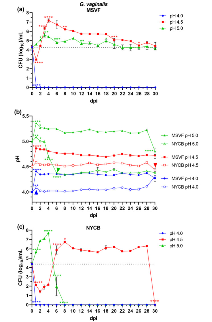
Fig. 1.
Growth patterns and changes in pH of G. vaginalis JCP8151A grown in MSVF and NYCB. G. vaginalis was grown overnight in NYCB at pH 7.3; cells were pelleted, washed, and resuspended in MSVF at pH 4.0, pH 4.5, and pH 5.0. One-mL aliquots were pipetted into the wells of a 24-well microtiter plate and inoculated with ~ 104 CFU. The plates were sealed with breathable membrane and the cultures were incubated at 37 °C under 5% CO2 in a humid chamber for 30 dpi. Samples were taken at 1-d intervals through 4 dpi and then every 2 d over the 30-d growth cycle and the CFU/mL and pH were determined. (a) Viability of G. vaginalis grown in MSVF for 30 d at starting pH of 4.0, 4.5, and 5.0; (b) pH of MSVF and NYCB at each time point throughout the growth cycle; (c) viability of G. vaginalis grown in NYCB for 30 d at starting pH of 4.0, 4.5, and 5.0. Each symbol represents the mean of three independent experiments ± SOM. Dotted lines (a, c) indicate starting CFU/mL. Arrowheads (b) indicate loss of viability. Two-way ANOVA with Tukey’s multiple comparisons posttest was done to determine significant differences between time points across the growth curves. *, P < 0.05; **, P < 0.01; ***, P < 0.001; ****, P < 0.0001

