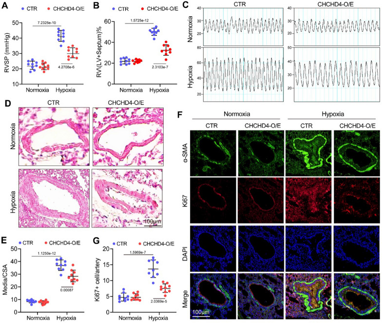
Fig. 3.
CHCHD4 attenuates hypoxic PAH. Hemodynamic examination was analyzed after the Normoxia or hypoxia exposure 28th day and then animals sacrificed. A The RVSP of the Normoxia and hypoxia rats with or without AAV-CHCHD4 treatment (n = 9). B Summary data of the right ventricle/(left ventricle + septum) weight ratio (n = 9). C Representative images of RVSP measurements in Normoxia and hypoxia rats with or without AAV-CHCHD4 treatment (n = 9). D H&E staining of pulmonary arteries in lung tissues in indicated animals. E Quantification of ratio of pulmonary arterial medial thickness to total vessel size (media/CSA) in each group (n = 9). F Double immunofluorescence staining of α-SMA (green) and Ki67 (red) in pulmonary arteries of the Normoxia and hypoxia rats with or without AAV-CHCHD4 treatment. G Quantification of Ki67 positive cells in pulmonary arteries (n = 9). Data are shown as the mean ± SEM. P value is showed in each figure

