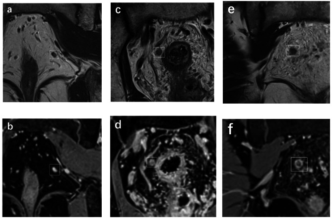
Fig. 4.
Coronal non-fat-suppressed high-spatial-resolution T2-weighted imaging (a, c and e); coronal fat-suppressed isotropic contrast-enhanced three-dimensional high-spatial-resolution T1-weighted imaging (b, d and f). The lymph nodes were signed by white boxes. a-b, Benign node with 3.7 mm in short-axis diameter, showed oval, smooth border, homogeneous signal and homogeneous enhancement. c-d, Benign node with 5.6 mm in short-axis diameter, showed round, irregular border, heterogeneous signal and heterogeneous enhancement. e-f, Malignant node with 5.8 mm in short-axis diameter, showed round, irregular border, heterogeneous signal and heterogeneous enhancement

