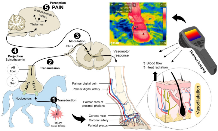
Figure 1.
Nociceptive pathway during laminitis. The inflammatory process and tissue injury caused by laminitis trigger the first phase of nociception. (1) Transduction. The noxious stimulus is recognized and transformed into an electrical signal by peripheral nerves (Aδ and C fibers), known as nociceptors. In these free nerve endings, receptors such as ASIC3 or TRPV1, among others, are activated to create action potentials that will be transmitted to the DRG of the spinal cord. (2) Transmission. Through Aδ and C fibers, the noxious input is transmitted to the spinal cord, to synapse with second-order neurons in the gray matter of the structure. (3) Modulation. Once the signal reaches the spinal cord, spinal interneurons are responsible for projecting or modulating the signal by releasing inhibitory or excitatory neurotransmitters. (4) Projection. Through the spinothalamic tract, the electrical signal reaches superior centers in the brain, mainly the thalamus. (5) Perception. From the thalamus, third-order neurons project to the somatosensory cortex, where the conscious recognition of pain is developed. Due to the interaction of the thalamus with other regions such as the hypothalamus, pain activates sympathetic centers, which causes physiological, endocrine, and behavioral responses. The vasomotor response, occurring as a result of pain and inflammation, causes vasodilation in the injured site to promote immune cells invasion to the injury site, with consequent healing. The increase in blood flow in the region also increases the amount of radiated heat from the skin. This element is captured by thermal cameras, helping to identify an inflammatory process in animals. ASIC: acid-sensing ion channel; DRG: dorsal root ganglion; TRPV1: transient potential receptor vanilloid 1.

