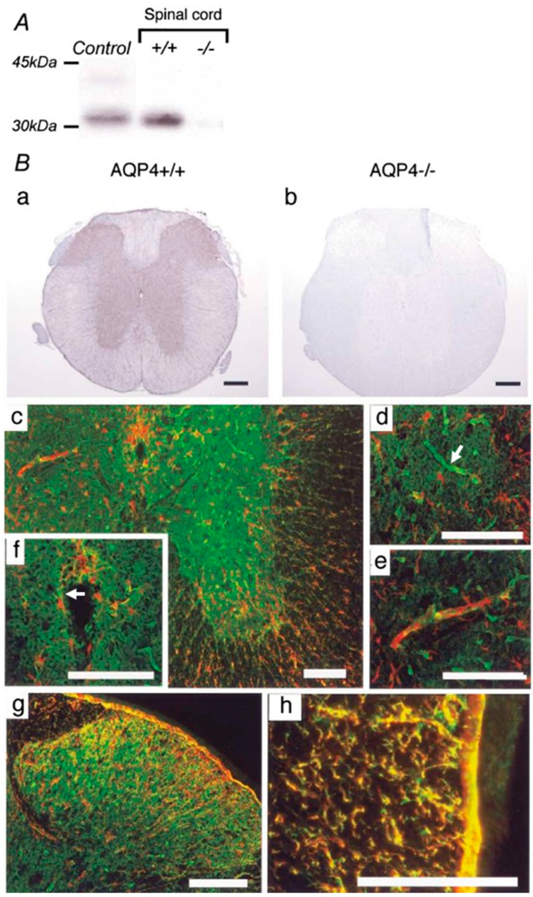
Figure 2.
AQP4 expression in mouse spinal cord. (A) Western blot analysis demonstrates an approximately 32 kDa band in control cerebral cortex (Control) and wild-type spinal cord (+/+). No expression was detected in the spinal cords of AQP4-deficient mice (−/−). (B) Immunohistochemistry for AQP4 reveals extensive expression in gray and white matter in the cervical spinal cords of wild-type mice (+/+) (a). No specific immunostaining was found in the spinal cords of AQP4 −/− mice (−/−). (b). Dense AQP4 staining (green) appeared extensively in gray matter (c), especially in astrocytic endfeet surrounding capillaries ((d), white arrow). GFAP-immunopositive (red) glial processes extending to capillaries were observed (e). Faint AQP4 and GFAP staining was detected in ependymal cells lining the central canal ((f), white arrow). GFAP and AQP4 were co-localized in fibrous thin astrocytes in the superficial dorsal horn (g). In white matter, AQP4 was co-expressed prominently with GFAP-immunoreactive radial fibrous glial processes surrounding the blood vessels and the glia limitans (h). ((c–h); red: GFAP, green: AQP4). Black scale bar = 0.2 mm. White scale bar = 0.1 mm. Figure and figure legend adopted from [49] with permission.

