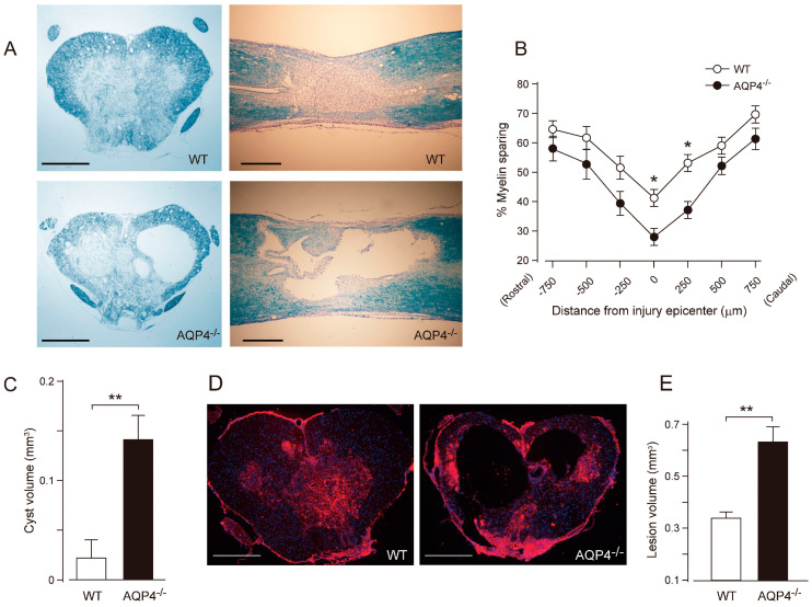
Figure 3.
Greater tissue damage and prominent cyst formation in AQP4−/− mice following a thoracic contusion injury. (A) Representative images of Luxol Fast Blue (LFB) staining at 42 days post-injury (dpi) (upper left, WT cross section; upper right, WT longitudinal section; lower left, AQP4−/− cross section, lower right; AQP4−/− longitudinal section). Note the greater demyelination and prominent cyst formation in AQP4−/− mice. (B) Stereological quantification of LFB staining shows significantly increased myelin loss in AQP4−/− mice. Data are represented as mean ± SEM, n = 7 each group; statistical significance was evaluated using a two-way ANOVA with Bonferroni post-hoc test, * p < 0.05. (C) Quantification of cyst volume shows significantly greater cyst volume in AQP4−/− mice (black bar) compared with WT (white bar). Data are represented as mean ± SEM, n = 7 each group; statistical comparisons were made using a Student’s t-test, ** p < 0.01. (D) Representative images of fibronectin staining at the injury epicenter (red, fibronectin; blue, DAPI; left, WT; right, AQP4−/−). (E) Stereological quantification of lesion volume delineated by fibronectin shows significantly greater lesion volume in AQP4−/− mice (black bar) compared with WT mice (white bar). Data are represented as mean ± SEM, n = 7 each group; statistical comparisons were made using a Student’s t-test, ** p < 0.01. Scale bars: A, D, 400 µm. Figure and figure legend adopted from [90] with permission.

