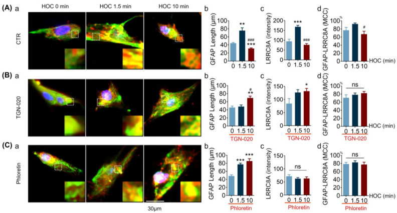
Figure 9.
TGN-20 or phloretin cause distinct effects on HOC-evoked GFAP filament length, LRRC8A expression, and GFAP-LRRC8A colocalization in cultured hypothalamic astrocytes. (A–C). Representative fluorescence images of GFAP (green), LRRC8A (red), and nuclei (blue, DAPI) at 0, 1.5, and 10 min HOC (a) in CTR ((A); n = 20, 15, and 10, respectively), 10 μmol/L of TGN-020 ((B); n = 17, 26, and 23, respectively), or 30 μmol/L of phloretin ((C); n = 20, 39, and 29, respectively). GFAP filament length (b), expression, i.e., staining/fluorescence intensity, of LRRC8A (c), and the MCC of GFAP with LRRC8A (d), respectively. Scale bar = 30 µm; *, p < 0.05, **; p < 0.01, ***, p < 0.005 compared with 0 min HOC; #, p < 0.05, ###, p < 0.005 compared with 1.5 min HOC; ANOVA, (Bonferroni for (C)b; Sidak for (A)c, (B)c, and (C)c; Dunn’s for (A)b, (A)d, (B)b, (B)d, and (D)d). Other annotations as in Figure 5.

