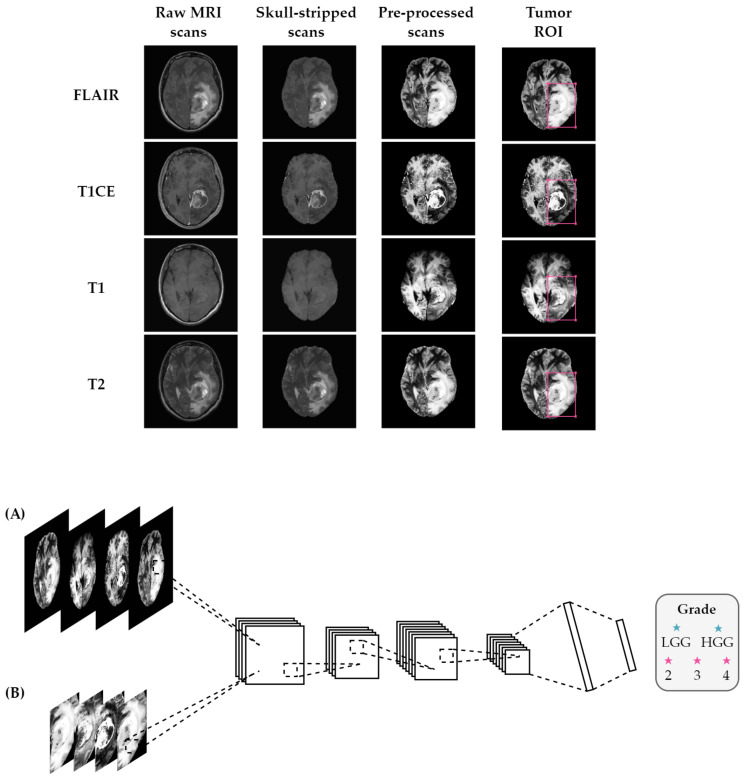
Figure 1.
Overview of the proposed method using (A) entire brain images and (B) tumor ROI. Before feeding the MRI scans into the classifier, the pipeline consisted of several pre-processing steps, including registration to a common atlas, skull-stripping, bias field correction, and normalization. The classifier takes FLAIR, T1 with contrast-enhancement, T1, and T2 scans stacked as input channels for the classification task.

