. 2023 Jun 15;59(57):8769–8778. doi: 10.1039/d3cc01682j

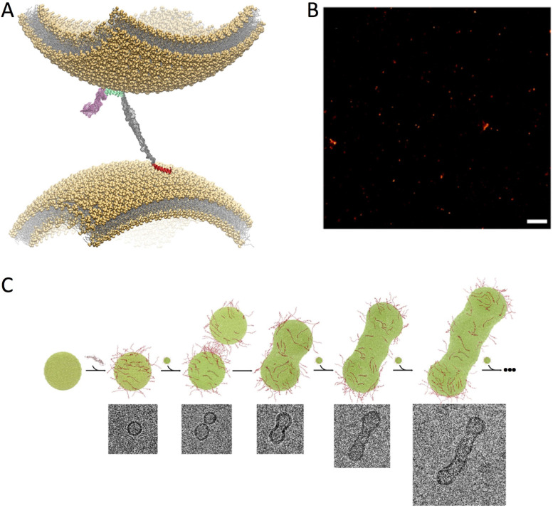
Fig. 3. Double-anchor mechanism. (A) Structural model of the double-anchor mechanism by which a single αS molecule binds simultaneously two lipid vesicles that are up to 150 Å apart, thereby promoting their indirect interaction. A first anchor, which spans residues 1 to 25 in N-terminal region (red), binds a vesicle in an amphipathic helical conformation while a second anchor, spanning the region 65 to 97 (cyan) of αS, binds a second vesicle. The C-terminal region (residues 99 to 140) and the linker (residues 26 to 59) are shown in pink and grey, respectively. (B) dSTORM imaging of the clustering of ex vivo SVs upon incubation in vitro with αS. Scale bar 1 μm. C) Stepwise representation of vesicle fusion promoted by αS in vitro as probed by cryo-EM. αS molecules (red) bind dynamically the surface of lipid vesicles (green) by promoting their fusion in a “worm-like” assembly. All images adapted from ref. 57, with permission from Nature Publishing Group, copyright 2016.

Fig. 3