. 2023 Jun 15;59(57):8769–8778. doi: 10.1039/d3cc01682j

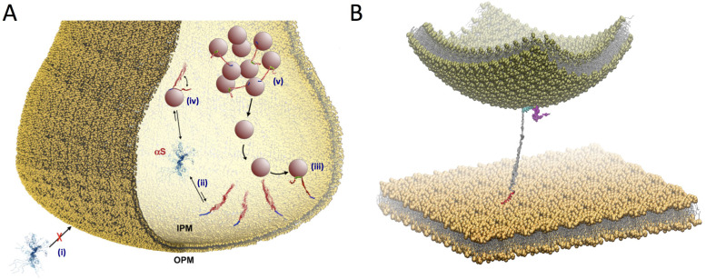
Fig. 4. Multiple membrane interactions by αS at the synaptic termini. (A) Scheme of competing interactions between αS and biological membranes at the presynaptic terminal. αS has negligible affinity to bind the outer PM (OPM) (i) but significantly higher affinity for the internal PM (IPM) (ii). The IPM bound state includes primarily the binding through the N-terminal anchor of αS (blue), with the region 65–140 (red) being mostly dissociated from the IPM surface. This conformation is optimal to promote a double-anchor mechanism (first anchor in blue and second anchor spanning residues 65–97 in green) to stabilise the SV docking on the IPM surface (iii). Competing with this process, the binding to SVs (iv) promotes the clustering of SV in pools (v) slowing down the SV diffusion toward the active zone. (B) Double anchor mechanism by which αS stabilizes SVs (green vesicle) on the IPM (flat yellow membrane) in a concentration dependent manner. The N-terminal region (red) binds the IPM while the central region (residues 65–97, cyan) interacts with the SV. The C-terminal region (residues 98–140) and the linker between the anchors (residues 26–64) are drawn in magenta and gray, respectively. Adapted from ref. 81, with permission from Nature Publishing Group, copyright 2021.

Fig. 4