. 2023 Jun 15;59(57):8769–8778. doi: 10.1039/d3cc01682j

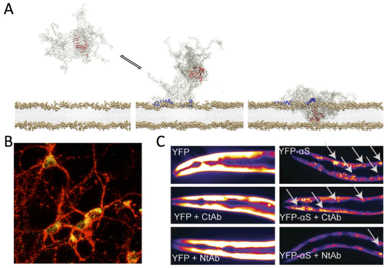
Fig. 5. Aberrant membrane disruption by αS oligomers. (A) Toxic type-B* αS oligomers were found to possess both structured (red) and disordered (grey) regions. The oligomers bind the membrane surface using the N-terminal regions of αS protomers in the assemblies (blue). The structured rigid core of the oligomers was found to insert into the lipid bilayer thereby disrupting its integrity. Adapted from ref. 1, with permission from the American Association for the Advancement of Science (licence number 5567500159295), copyright 2017. (B) Membrane interaction of type-B* αS oligomers with primary cortical neurons imaged using confocal scanning microscopy. Red and green fluorescence indicates the cell membranes and the αS oligomers, respectively. Adapted from ref. 132, with permission from American Chemical Society, copyright 2019. (C) Effect antibodies targeting the N- and C-terminal regions of αS (Nt-Ab and Ct-Ab) in C. elegans models of αS toxicity. These worms overexpress YFP-tagged αS in the muscles and the aggregation of the protein generates toxicity. Representative fluorescence microscopy images of worms at day 11 of adulthood expressing YFP (left) or YFP-αS (right) is shown in the absence (top) or presence of 0.4 μM of Ct-Ab (middle) or Nt-Ab (bottom). The inclusions are indicated by white arrows. Adapted from ref. 132, with permission from American Chemical Society, copyright 2019.

Fig. 5