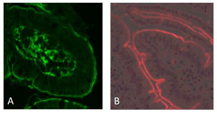
Figure 1.
Immunofluorescence staining of transglutaminase 2 (using mab CUB 7402, green fluorescence) and gliadin peptides (mab A161, red fluorescence) in a duodenal biopsy from a CeD patient. The majority of the total TG2 is localized near the basement membrane and in the lamina propria. The luminal surface of the villous epithelium is labeled with equally strong intensity (panel (A)). Gliadin peptides (33-mer, non-deamidated, and deamidated) also localize on the luminal surface of the villous epithelium (panel (B)). Original magnification: ×200.

