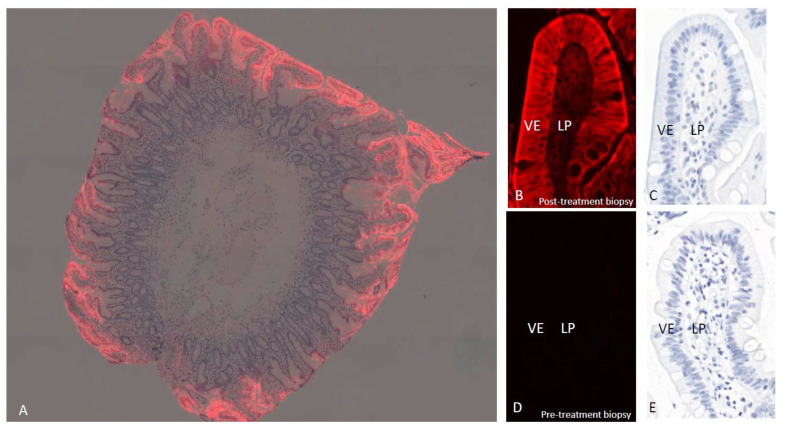
Figure 2.
Localization of ZED1227 by mab A083 immunofluorescence (red) in a duodenal biopsy from a ZED1227-treated patient at low magnification (panel (A), image blended with hematoxylin counterstain). The signal accumulation in the villous epithelium is evident. At higher magnification, a post-treatment (100 mg/day) biopsy shows the labeling localized mainly in the apical surface of the villous epithelium (VE) (panel (B), counterstain in panel (C)). Lower-intensity ZED1227-TG2 labeling can be seen in the lamina propria (LP). The fluorescence signal is absent in a pre-treatment biopsy from the same patient (panels (D,E)).

