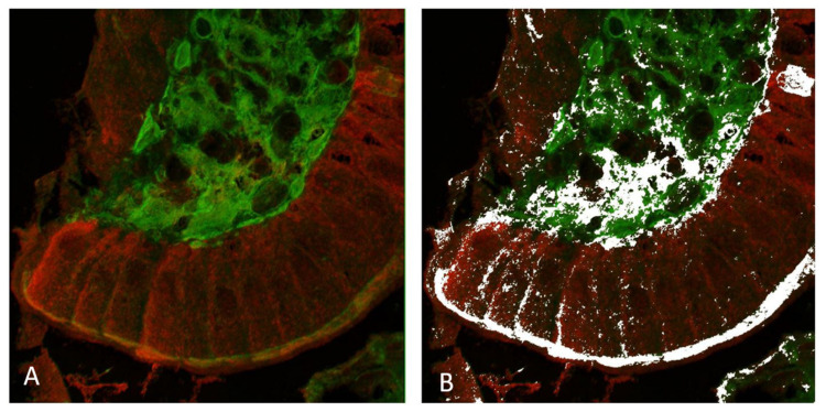
Figure 4.
Confocal microscopy imaging of total TG2 (mab CUB 7402, in green) and ZED1227 (mab A083, in red) by double immunofluorescence staining (panel (A)). White pixels in panel (B) show the areas with statistically significant co-localization defined by the ImageJ/FIJI image analysis co-localization algorithm. Original magnification: ×600.

