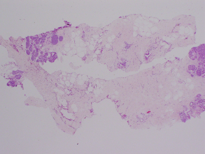
Figure 2b:

Images in 48-year-old woman with nipple discharge. (a) US scan shows small, irregular, hypoechoic mass (arrows), for which biopsy was recommended. (b) Low-power photomicrograph (H-E stain; original magnification, ×40) of core biopsy specimen reveals normal breast parenchyma with foci of ALH. This was discordant with imaging finding of mass, and surgical excision was recommended. (c) Photomicrograph of surgical specimen (H-E stain; original magnification, ×200) reveals small papilloma, which was fully excised.
