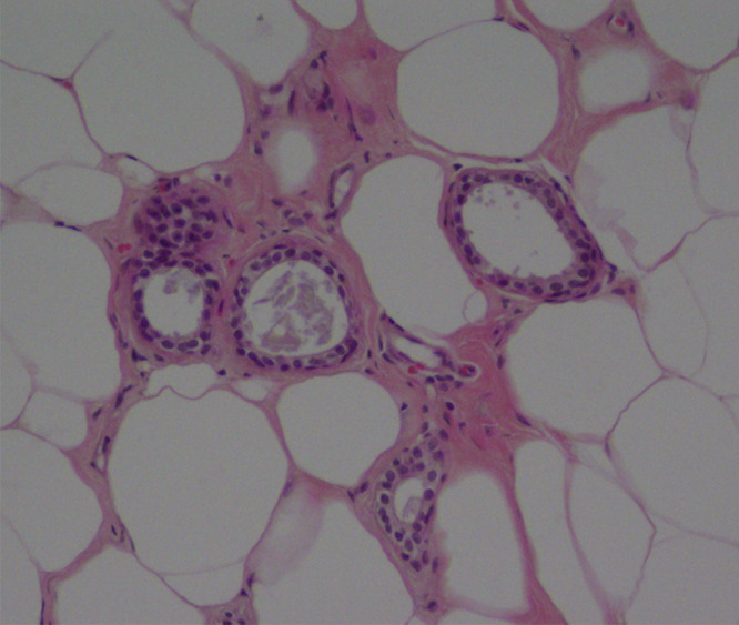
Figure 3c:

(a) Mammogram in 52-year-old woman shows suspicious fine linear branching microcalcifications (arrow), for which stereotaxic core biopsy was recommended and performed. (b) Radiograph of core specimen obtained at stereotaxic biopsy reveals numerous microcalcifications (circles). (c) Photomicrograph of core biopsy specimen (H-E stain; original magnification, ×200) reveals only scant benign calcification and foci of ALH (not pictured). The paucity of calcifications identified at histologic examination combined with the benign diagnosis was thought to be discordant, and surgical excision was recommended. (d) Photomicrograph of surgical specimen (H-E stain; original magnification, ×400) shows DCIS.
