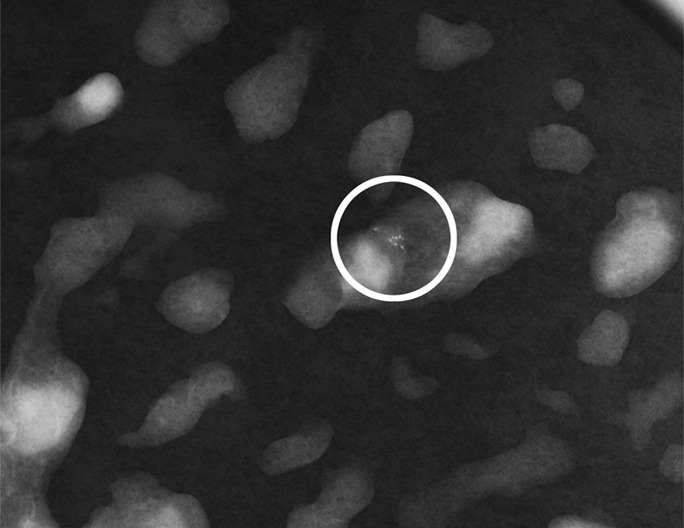
Figure 4b:

(a) Magnification view of left breast in 62-year-old woman reveals fine pleomorphic microcalcifications in segmental distribution (arrows), for which stereotaxic core biopsy was recommended. (b) Radiograph of specimen from stereotaxic biopsy reveals adequate sampling of suspicious microcalcifications (circle). (c) Photomicrograph of specimen (H-E stain; original magnification, ×400) reveals fibrocystic changes with scattered microcalcifications. (d) Although this histologic finding was concordant with imaging features, florid LCIS (H-E stain; original magnification, ×200) was also identified in core specimens and excision was recommended. Final excision showed noncalcified florid LCIS.
