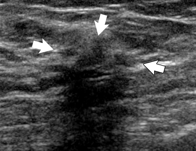
Figure 6a:

(a) Image in 54-year-old woman with irregular hypoechoic mass (arrows) with posterior acoustic shadowing, for which US-guided core biopsy was recommended. (b) Photomicrograph of core biopsy specimen (H-E stain; original magnification, ×40) shows extensive dense sclerosis in all cores (light pink stroma at left and top) and foci of ALH (right). Sclerosis was thought to be concordant with imaging features. Surgical excision revealed only ALH.
