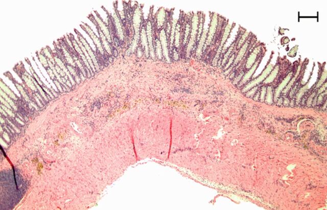
Figure 3e:
Representative (a, c, e) hematoxylin-eosin– and (b, d, f) Masson trichrome–stained histopathologic images with ×5 magnification. (a, b) Phosphate-buffered saline negative control rat colon is normal without bowel wall thickening, acute inflammation, or fibrosis. (c, d) Rat in acute inflammation cohort demonstrates bowel wall thickening with extensive acute inflammation, mucosal ulceration, and necrosis. (e, f) Fibrotic cohort rat demonstrates bowel wall thickening with transmural fibrosis (blue staining involving submucosa and muscularis propria).

