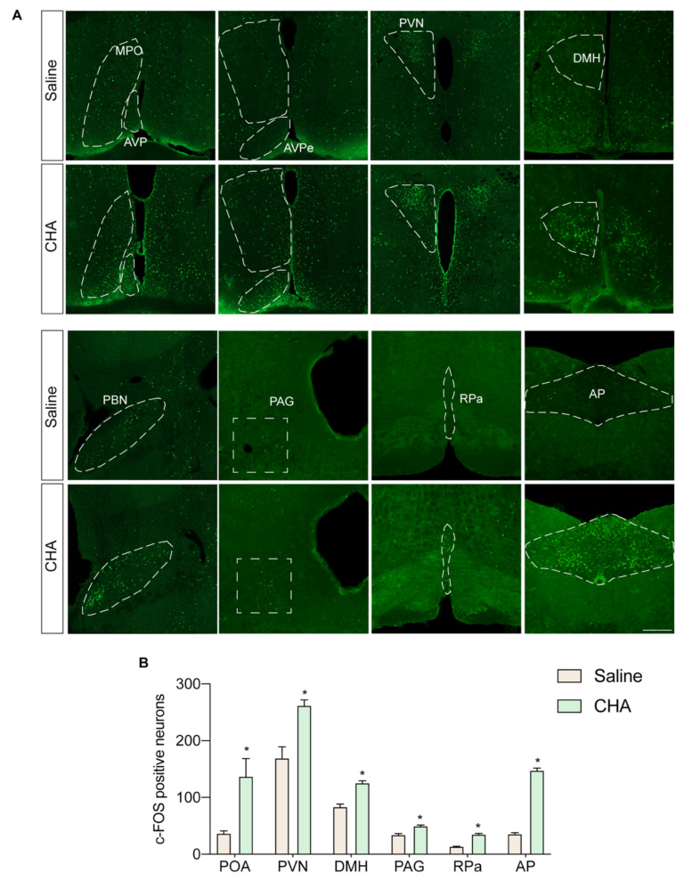
Figure 4.
Whole brain tracing of neural circuit activated by CHA. (A) c-Fos staining of POA, PVN, DMH, PBN, PAG, RPa, and AP nucleus. (B) Quantification of c-Fos-positive neurons in (A). POA: preoptic area; MPO: medial preoptic area; AVP: anterior ventral preoptic area; PVN: paraventricular nucleus; DMH: dorsal medial hypothalamus; PBN: parabrachial nucleus; PAG: periaqueductal gray; RPa: raphe pallidus; AP: area postrema. p < 0.05 for *. Scale bar = 200 µm.

