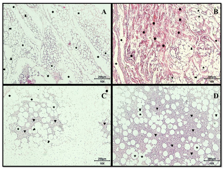
Figure 4.
Representative images of histological sections of adipose tissue from adult male Wistar rats (31 weeks), with inflammation induced by the HGLI diet, submitted to different treatments and evaluated after ten days of the experiment (11th day), stained with hematoxylin-eosin (HE) belonging to the following: (A) Untreated group. (B) Conventional treatment. (C) Test treatment 1. (D) Test treatment 2. Total magnification 100× (Objective Lens 10×), scale bar: 200 μm. Panoramic evidence of the presence of white adipose tissue consisting of numerous unilocular adipocytes ( ) primarily intact, showing focal areas of membrane destruction (
 ), extensive regions of lipolysis channels (
 ), vast areas of fibrosis consisting of dense non-patterned connective tissue in areas of adipocyte destruction (
 ), and the presence of the focal regions of multilocular adipocytes (
 ). No treatment: HGLI diet + 1 mL of water by gavage; conventional treatment: nutritionally adequate diet (Labina® feed) + 1 mL of water per gavage; test treatment 1: HGLI diet + 1 mL of CE at a concentration of 12.5 mg/kg by gavage; test treatment 2: HGLI diet + 1 mL of EPG at a concentration of 50 mg/kg by gavage; HGLI diet: mixture composed of Labina®, condensed milk and sugar (1:1:0.21 w/w/w); HGLI: high glycemic index and high glycemic load diet; CE: crude extract rich in carotenoids from Cantaloupe melons; EPG: crude extract rich in carotenoids from Cantaloupe melons nanoencapsulated in porcine gelatin.

