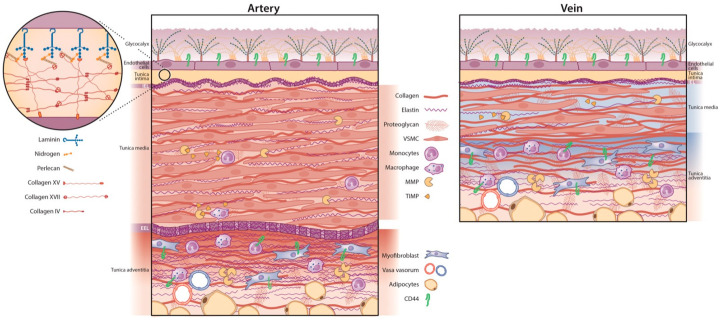
Figure 1.
Overview of the arterial versus venous vessel layers and their respective ECM. The luminal side of both vessel types contains an intimal layer with endothelial cells (ECs) covered by the glycocalyx. The basal lamina is a mesh-like structure situated underneath the ECs. The internal elastic lamina (IEL) separates the tunica intima and media. The arterial (left panel) tunica media is thicker and more elastic than its venous counterpart (right panel) with more vascular smooth muscle cells (VSMCs), organised with elastin into a contractile-elastic unit. Arteries have a prominent external elastic lamina (EEL). Veins are less muscular with a lower elastin-to-collagen ratio. The tunica adventitia contains collagen-producing (myo) fibroblasts, surrounded by perivascular adipose tissue and the vasa vasorum: a capillary network of minor blood vessels. Matrix metalloproteinases (MMPs) are proteinases that regulate ECM degradation and extend into the media and adventitia. They are inhibited by tissue inhibitors of metalloproteinases (TIMPs). CD44 is a cell-surface glycoprotein receptor found in the venous and arterial adventitial layer and venous tunica intima. CD44 is expressed by ECs and can bind ECM components, such as collagen, fibronectin, MMPs, and hyaluronic acid. ECM = extracellular matrix.

