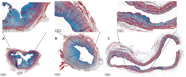
Figure 3.
Differential ECM deposition in human AVFs. Samples were stained with Masson Trichrome. ECM is shown in blue, and smooth muscle cells are stained red. Scale bars are 100 µm in the bottom images and 500 µm in the inlay. (A) shows an AVF with little outward remodelling and a lot of ECM deposition in the IH, as shown in the inlay, indicative of a fibrotic AVF. (B) AVF with wall thickening and some ECM deposition amongst well-organised muscle fibres. (C) AVF with excessive OR and little ECM deposition, indicative of aneurysm formation. AVF = arteriovenous fistula, ECM = extracellular matrix, IH = intimal hyperplasia, OR = outward remodelling.

