Figure 2.
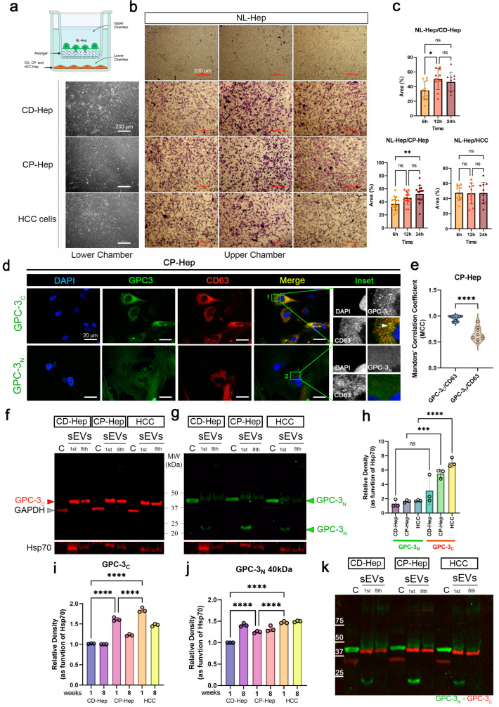
C-terminal GPC-3 domain is localized in hepatocyte small EVs. (a) schematic of the multichambered system. Normal hepatocytes plated in the upper chamber coated with Matrigel and CD-, CP- and HCC hepatocytes plated in the lower chamber. (b) Representative phase contrast images of the lower chamber (left column) and matrigel chamber (upper chamber) with migrating hepatocytes (magnification 4×, scale bar: 200 µm). (c) Quantification of the area (expressed in % of covered area) covered by migrating cells in NL-Hep/CD-Hep, NL-Hep/CP-Hep and NL-Hep/HCC systems. Ordinary one-way ANOVA and Tukey’s multiple comparison test was performed, *, p < 0.038 and **, p < 0.017. (d) Representative IF images of CP-Hep stained with DAPI-nuclei (blue). GPC-3C and GPC-3N (green), CD63 (red) and merged channels. Inset of magnified region of interests are reported (magnification 63×, scale bar: 20 µm). (e) Manders’ Colocalization Coefficients are reported to measure colocalization of GPC-3 domains in CD63 positive sEVs (unpaired t-test ****, p < 0.001). (f) Immunoblot of GPC-3C and GPC-3N (g) in sEVs isolated from CD-, CP- and HCC-Hep at 1st and 8th week of cultures, alongside cytoplasmic fractions. GAPDH, loading control for cytoplasm, Hsp70 as loading control for sEVs. (h) Relative density of GPC-3N and GPC-3C in sEVs fractions (Ordinary one-way ANOVA, Tukey’s multiple comparison test ***, p < 0.001 and, ****), (i) Relative density of GPC-3C in sEVs fractions in 1 and 8 weeks (Ordinary one-way ANOVA, Tukey’s multiple comparison test, ****). (j) Relative density of GPC-3N in sEVs fractions in 1 and 8 weeks (Ordinary one-way ANOVA, Tukey’s multiple comparison test, ****). (k) IB overlapped GPC-3C (red) and GPC-3N (green).

