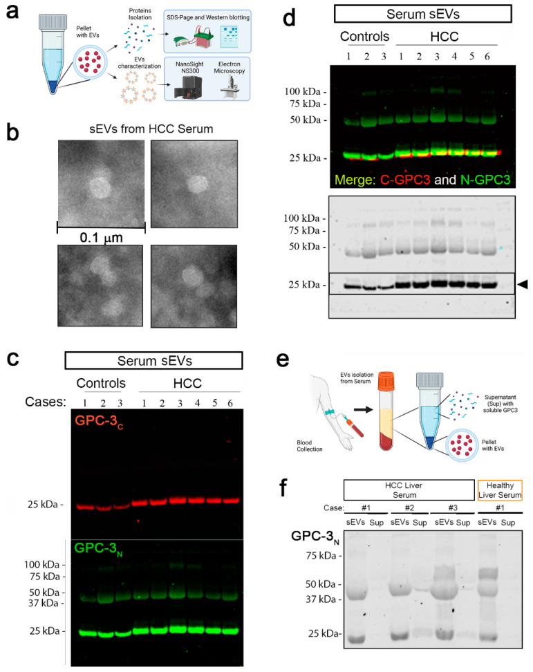
Figure 4.
Glypican-3 in human serum small EVs. (a) Schematic representation of sEVs extraction. Pellets were used for size and morphology characterization using Nano Sight NS3000 and TEM, respectively. Protein contents were evaluated with western blot. (b) Representative TEM image of sEVs isolated from HCC serum (scale bar: 0.1 µm). (c) GPC-3 sEVs content was evaluated by WB in healthy controls and HCC cases. GPC-3C (red-top panel) and GPC-3N (green-bottom panel) antibodies were used for immunoblotting. (d) Upper blot—Merged immunoblot of N- and C-terminal GPC-3 IBs, Lower Blot—GPC-3N IB is represented as well in black/white. (e) schematic representation for serum sEVs and supernatant isolation to verify GPC-3 soluble content. (f) IB of GPC-3N in sEVs and corresponding supernatant (Sup) in three HCC cases and 1 control.

