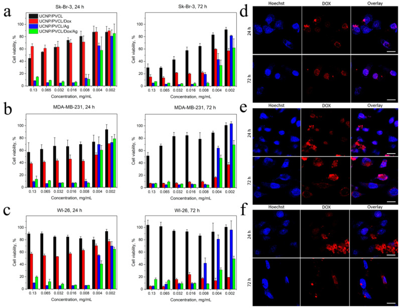
Figure 3.
Cell viability of (a) human breast adenocarcinoma Sk-Br-3 cells. (b) Human breast adenocarcinoma MDA-MB-231 cells. (c) Human WI-26 fibroblasts under NP treatment, 24 h and 72 h incubation, MTT assay. The data are presented as mean ± SD. Confocal microscopy of (d) Sk-Br-3 cells, (e) MDA-MB-231 cells, (f) WI-26 fibroblasts incubated with PMAO–PVCL Dox nanocomplexes within 24 h and 72 h. Blue is for Hoechst 33342 (cell nuclei), red is for Dox. Scale bar is 20 µm.

