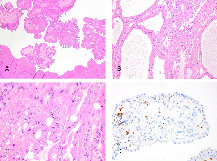
Figure 2.

FH-deficient RCCs usually presents with a variable morphologic patterns that frequently include papillary and intracystic (A) and tubulocystic growth (B). Prominent “cherry-red” nucleoli are at least focally present (C). FH immunostaining is absent in the tumour cells (D).
