Figure 4.

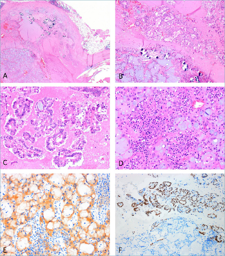
ALK-rearranged RCC has typically heterogeneous morphology. (A) A circumscribed tumour with peripheral pseudocapsule seen at low power, with easily recognisable pools of mucin and aggregates of psammoma bodies/calcifications. (B) At higher power, papillary formations can be seen (top), adjacent to tubules with luminal mucin (bottom). (C) Papillary formations are set in a necrotic background. (D) Foci of mucin-containing signet-ring cells with an inflammatory background were also present. (E) ALK immunostaining is positive in the neoplastic cells. (F) Papillary formations also demonstrated unusual nuclear immunoreactivity for TTF1 (thyroglobulin was negative, not shown).
